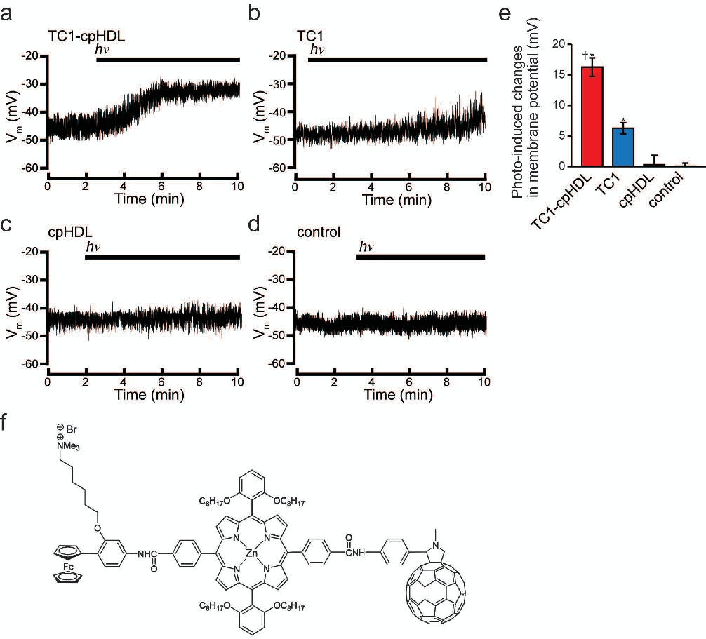
Fig. 1. Photo-induced depolarization of PC12 cells containing TC1. Representative traces of time-dependent changes in the membrane potential (Vm) under illumination (black bar, hν: 525−550 nm, input power 2 mW cm−2) are shown for PC12 cells treated for 3 min with (a) TC1-loaded cell-penetrating high-density lipoprotein (TC1-cpHDL), (b) TC1, (c) cpHDL, (d) DMSO (control) and then washed with a Tyrode bath solution. (e) The average of the photo-induced changes in the membrane potential (mV) in TC1-cpHDL, TC1, cpHDL, and neither TC1 or cpHDL (control). (f) Molecular structure of TC1. TC1 is photo-induced charge-separation molecule consisting of ferrocene-zincporphyrin-fullerene (Fc-ZnP-C60) triads (see Materials and Methods for details). *p<0.05 vs. control, †p<0.05 vs. TC1 (n = 5-7). hν: photon energy where h is Planck's constant 6.626×10⁻³⁴, ν is frequency.